IIT researchers take rare look inside foetal brain — one slice at a time, The foetal brain atlas is the largest of its kind and can detect brain disorders like autism.
Researchers at IIT Madras have unveiled a cutting-edge tool — a detailed 3D map of five developing baby brains from the second trimester.
This map, now the most detailed high-resolution 3D representation of the foetal brain, shows how it undergoes rapid growth during this critical stage and can detect possibilities of brain disorders like autism.
Called DHARINI, this brain atlas is the largest of its kind and the only one that captures the developing brain at such an early stage. It uses advanced technology to map over 5,000 brain sections and more than 500 brain regions. It will be completely free for anyone to access, opening up new possibilities for understanding how our brains grow.

Why is this brain map significant?
“This is groundbreaking research for clinicians — it will help us study how the human brain develops in the womb.
This data could provide crucial insights into developmental disorders like autism, which remain poorly understood and managed. “It may also help explain why some children suffer permanent damage and develop cerebral palsy after hypoxia (a lack of oxygen) while others recover without lasting effects,”.
Additionally, the findings could shed light on changes in the adult brain linked to mental health conditions such as depression or bipolar disorder.
“The output from this will keep scientists busy around the world for years to come. The developments in artificial intelligence and machine learning came from us wondering about how the brain works and trying to recreate that magic using silicon.
Better understanding of human brains will create newer models, better models.
And though AI is being talked about at the moment, there are improvements that need to be made.
The brain atlas created by IIT Madras is not only the largest dataset in the world, it is also the only one that has been able to capture the growing brain in foetuses. The only other publicly available brain atlas such as this was released by US Allen Institute for Brain Science in 2016. It captured the brain of an adult woman in 1,356 plates.
How was the mapping done?
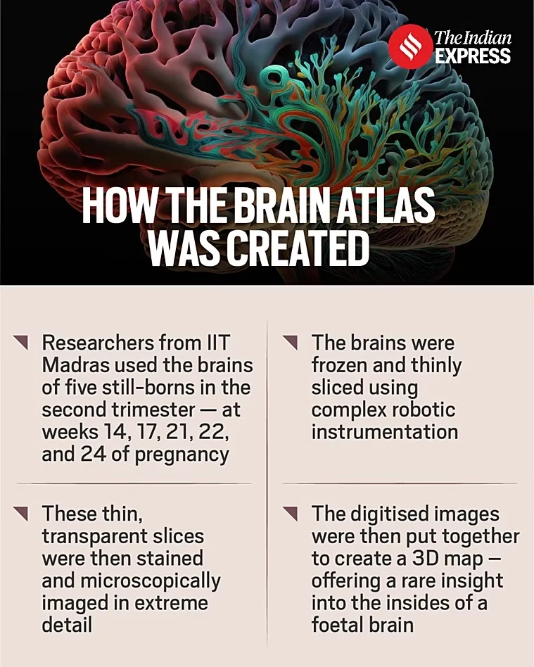
To capture the complex structures of the brain at a cellular level, researchers from IIT Madras used the brains of five still-borns in the second trimester — at 14, 17, 21, 22 and 24 weeks of pregnancy.
The brains were frozen and thinly sliced, enabling scientists to see the structures.
“The brains are very thinly sliced using complex robotic instrumentation — the slices are of just 10 to 20 micron thickness which is equivalent to 1/10th or 1/5th the thickness of human hair”.
These thin slices, which become transparent, are then stained and microscopically imaged in extreme detail. Once digitised, these slides are put together to create a 3D map.
The technology used for freezing, slicing, creating plates, digitising, and putting the map together has been indigenously developed by the IIT researchers.
The IIT centre has collected nearly 230 brains of still- borns and neonates as of now. The institute also plans to study paediatric and older brains.
Source: Indian Express

